Comments, observations and thoughts from two bloggers on applied statistics, higher education and epidemiology. Joseph is an associate professor. Mark is a professional statistician and former math teacher.
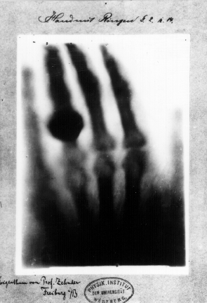
Hand mit Ringen (Hand with Rings): print of Wilhelm Röntgen's
first "medical" X-ray, of his wife's hand, taken on 22 December 1895 and
presented to Ludwig Zehnder of the Physik Institut, University of Freiburg, on 1 January 1896
Conventional wisdom has it that our modern pace of technological change is so fast that someone from 100 years or so ago would find it incomprehensible. I don't buy that for at least a couple of reasons. First, we have a tendency to forget just how long some technologies are taking to get here (do you have any idea how long autonomous vehicles have been just around the corner?). Second, we tend to grossly underestimate how quickly many technologies of the past were disseminated.
For the canonical example of rapid adoption, check out the following excerpts from the Wikipedia article on the history of the x-ray starting with Röntgen's breakthrough, followed by some excerpts from Scientific American. Pay close attention to the timeline and keep in mind, this is how people reacted to a technology so new and unexpected that it bordered on unimaginable.
[Emphasis added for dates throughout.]
On8 November 1895, German physics professor Wilhelm Röntgen stumbled on X-rays while experimenting with Lenard tubes and Crookes tubes
and began studying them. He wrote an initial report "On a new kind of
ray: A preliminary communication" and on 28 December 1895, submitted it
to Würzburg's Physical-Medical Society journal.
...
The discovery of X-rays generated significant interest. Röntgen's biographer Otto Glasser estimated that, in 1896 alone, as many as 49 essays and 1044 articles about the new rays were published.[25]
This was probably a conservative estimate, if one considers that nearly
every paper around the world extensively reported about the new
discovery, with a magazine such as Science dedicating as many as 23 articles to it in that year alone.
...
Röntgen immediately noticed X-rays could have medical applications.
Along with his 28 December Physical-Medical Society submission, he sent a
letter to physicians he knew around Europe (1 January 1896).[29] News (and the creation of "shadowgrams") spread rapidly with Scottish electrical engineer Alan Archibald Campbell-Swinton
being the first after Röntgen to create an X-ray (of a hand). Through
February, there were 46 experimenters taking up the technique in North
America alone.[29]
The first use of X-rays under clinical conditions was by John Hall-Edwards
in Birmingham, England on 11 January 1896, when he radiographed a
needle stuck in the hand of an associate. On 14 February 1896,
Hall-Edwards was also the first to use X-rays in a surgical operation.[30]
Images by James Green, from "Sciagraphs of British Batrachians and Reptiles" (1897), featuring (from left) Rana esculenta (now Pelophylax lessonae), Lacerta vivipara (now Zootoca vivipara), and Lacerta agilis
In early 1896, several weeks after Röntgen's discovery, Ivan Romanovich Tarkhanov irradiated frogs and insects with X-rays, concluding that the rays "not only photograph, but also affect the living function".[31] At around the same time, the zoological illustrator James Green began to use X-rays to examine fragile specimens. George Albert Boulenger first mentioned this work in a paper he delivered before the Zoological Society of London in May 1896. The book Sciagraphs of British Batrachians and Reptiles
(sciagraph is an obsolete name for an X-ray photograph), by Green and
James H. Gardiner, with a foreword by Boulenger, was published in 1897.[32][33]
The first medical X-ray made in the United States was obtained
using a discharge tube of Pului's design. In January 1896, on reading of
Röntgen's discovery, Frank Austin of Dartmouth College
tested all of the discharge tubes in the physics laboratory and found
that only the Pului tube produced X-rays. This was a result of Pului's
inclusion of an oblique "target" of mica, used for holding samples of fluorescent
material, within the tube. On 3 February 1896, Gilman Frost, professor
of medicine at the college, and his brother Edwin Frost, professor of
physics, exposed the wrist of Eddie McCarthy, whom Gilman had treated
some weeks earlier for a fracture, to the X-rays and collected the
resulting image of the broken bone on gelatin photographic plates obtained from Howard Langill, a local photographer also interested in Röntgen's work.


 And to get a feel for the impact on the popular imagination.